The MitoPedia terminology is developed continuously in the spirit of Gentle Science.
»O2k-Publications: O2k-Fluorometry
»Discussion: Fluorometry, »Respirometry, »Spectrophotometry
| Term | Abbreviation | Description |
|---|---|---|
| Absorbance | A | Also known as attenuation or extinction, absorbance (A) is a measure of the difference between the incident light intensity (I0) and the intensity of light emerging from a sample (I). It is defined as: A = log (I0/I) |
| Amp calibration - DatLab | F5 | Amp calibration indicates the calibration of the amperometric O2k-channel. |
| Amplex UltraRed | AmR | Amplex® UltraRed (AmR) is used as an extrinsic fluorophore for measurement of hydrogen peroxide production (ROS) by cells or mitochondrial preparations. The reaction of H2O2 and AmR is catalyzed by horseradish peroxidase to produce the red fluorescent compound resorufin (excitation wavelength 563 nm, emission 587 nm; the fluorescent product according to the supplier is called UltroxRed in the case of Amplex® UltraRed which has a similar structure to resorufin). The change of emitted fluorescence intensity is directly proportional to the concentration of H2O2 added, whereby the H2O2 is consumed. |
| Averaging | In order to improve the signal-to-noise ratio a number of sequential spectra may be averaged over time. The number of spectra to be averaged can be set prior to carrying out the measurements, or afterwards during data analysis. | |
| Bandwidth | Bandwidth is measured in nanometers in terms of the full width half maximum of a peak. This is the portion of the peak that is greater than half of the maximum intensity of that peak. | |
| Blank | In fluorometry and transmission spectrophotometry blank cuvettes (with no samples in them) are used to carry out the balance. | |
| Calcium | Ca | Ca2+ is a major signaling molecule in both prokaryotes and eukaryotes. Its cytoplasmic concentration is tightly regulated by transporters in the plasma membrane and in the membranes of various organelles. For this purpose, it is either extruded from the cell through exchangers and pumps or stored in organelles such as the endoplasmic reticulum and the mitochondria. Changes in the concentration of the cation regulate numerous enzymes including many involved in ATP utilizing and in ATP generating pathways and thus ultimately control metabolic activity of mitochondria and of the entire cell. Measuring changes in Ca2+ levels is thus of considerable interest in the context of high-resolution respirometry. |
| Calcium Green | CaG | Calcium GreenTM (CaG) denotes a family of extrinsic fluorophores applied for measurement of Ca2+ concentration with mitochondrial preparations. This dye fluoresces when bound to Ca2+. When measuring mitochondrial calcium uptake it is possible to observe the increase of the CaG signal upon calcium titration, followed by the decrease of CaG signal due to the uptake. |
| Carboxy SNARF 1 | SNARF | Carboxy SNARF® 1 is a cell-impermeant pH indicator dye. The pKa of ~7.5 makes it useful for measuring pH in the range of pH 7 to pH 8. The emission shifts from yellow-orange at low pH to deep red fluorescence at high pH. Ratiometric fluorometry, therefore, is applied at two emission wavelengths,such as 580 nm and 640 nm. Relative molecular mass: Mr = 453.45 |
| Cuvettes | Cuvettes are used in fluorometry and transmission spectrophotometry to contain the samples. Use of the term 'cells' for cuvettes is discouraged, to avoid confusion with 'living cells'. Traditionally cuvettes have a square cross-section (10 x 10 mm). For many applications they are made of transparent plastic. Glass cells are used where samples may contain plastic solvents, and for some applications requiring measurements below 300 nm, quartz glass or high purity fused silica cuvettes may be necessary. | |
| DTPA | DTPA | DTPA (Diethylenetriamine-N,N,N',N,N-pentaacetic acid, pentetic acid,(Carboxymethyl)imino]bis(ethylenenitrilo)-tetra-acetic acid) is a polyaminopolycarboxylic acid (like EDTA) chelator of metal cations. DTPA wraps around a metal ion by forming up to eight bounds, because each COO- group and and N-center serves a center for chelation. With transition metals the number of bounds is less than eight. The compound is not cell membrane permeable. In general, it chelates multivalent ions stronger than EDTA. |
| Detector | A detector is a device that converts the light falling upon it into a current or voltage that is proportional to the light intensity. The most common devices in current use for fluorometry and spectrophotometry are photodiodes and photodiode arrays. | |
| Diffraction gratings | Diffraction gratings are dispersion devices that are made from glass etched with fine grooves, spaced at the same order of magnitude as the wavelength of the light to be dispersed, and then coated with aluminium to reflect the light to the photodiode array. Diffraction gratings reflect the light in different orders and filters need to be incorporated to prevent overlapping. | |
| Dilution effect | Dilution of the concentration of a compound or sample in the experimental chamber by a titration of another solution into the chamber. | |
| Dispersion devices | A dispersion device diffracts light at different angles according to its wavelength. Traditionally, prisms and diffraction gratings are used, the latter now being the most commonly used device in a spectrofluorometer or spectrophotometer. | |
| Drift | The most common cause of drift is variation in the intensity of the light source. The effect of this can be minimised by carrying out a balance at frequent intervals. | |
| Electron leak | Electrons that escape the electron transfer pathway without completing the reduction of oxygen to water at cytochrome c oxidase, causing the production of ROS. The rate of electron leak depends on the topology of the complex, the redox state of the moiety responsible of electron leakiness and usually on the protonmotive force (Δp). In some cases, the Δp dependance relies more on the ∆pH component than in the ∆Ψ. | |
| Extrinsic fluorophores | Extrinsic fluorophores are molecules labelled with a fluorescent dye (as opposed to intrinsic fluorescence or autofluorescence of molecules which does not require such labelling). They are available for a wide range of parameters including ROS (H2O2, Amplex red) (HOO-, MitoSOX) , mitochondrial membrane potential (Safranin, JC1, TMRM, Rhodamine 123), Ca2+ (Fura2, Indo 1, Calcium Green), pH (Fluorescein, HPTS, SNAFL-1), Mg2+ (Magnesium Green) and redox state (roGFP). | |
| Filters | Filters are materials that have wavelength-dependent transmission characteristics. They are can be used to select the wavelength range of the light emerging from a light source, or the range entering the detector, having passed through the sample. In particular they are used in fluorometry to exclude wavelengths greater than the excitation wavelength from reaching the sample, preventing absorption interfering with the emitted fluorescence. Standard filters can also be used for calibrating purposes. | |
| Fluorescence | Fluorescence is the name given to light emitted by a substance when it is illuminated (excited) by light at a shorter wavelength. The incident light causes an electron transition to a higher energy band in the molecules. The electron then spontaneously returns to its original energy state emitting a photon. The intensity of the emitted light is proportional to the concentration of the substance. Fluorescence is one form of Luminescence, especially Photoluminescence. | |
| Fluorescent marker | See Extrinsic fluorophores | |
| Fluorometric dyes | Extrinsic fluorophores; fluorescent markers. | |
| Fluorometry | Fluorometry (or fluorimetry) is the general term given to the method of measuring the fluorescent emission of a substance following excitation by light at a shorter wavelength. | |
| Fluorophore | A fluorophore is a fluorescent substance that may occur naturally (intrinsic fluorophores) or that may be added to a sample or preparation whereby the fluorescence intensity is proportional to the concentration of a specific species or parameter within the sample. These are extrinsic fluorophores, also referred to as fluorescent markers. | |
| Flux / Slope | J | Flux / Slope is the time derivative of the signal. In DatLab, Flux / Slope is the name of the pull-down menu for (1) normalization of flux (chamber volume-specific flux, sample-specific flux or flow, or flux control ratios), (2) flux baseline correction, (3) Instrumental background oxygen flux, and (4) flux smoothing, selection of the scaling factor, and stoichiometric normalization using a stoichiometric coefficient. Before changing the normalization of flux from volume-specific flux to sample-specific flux or flow, or flux control ratios, please be sure to use the standard Layout 04a (Flux per volume) or 04b (Flux per volume overlay). When starting with the instrumental standard Layouts 1-3, which display the O2 slope negative, the sample-specific flux or flow, or flux control ratios will not be automatically background corrected. To obtain the background corrected specific flux or flux control ratios, it is needed to tick the background correction in the lower part of the slope configuration window. Background correction is especially critical when performing measurements in a high oxygen regime or using samples with a low respiratory flux or flow. |
| Flux baseline correction | bc | Flux baseline correction provides the option to display the plot and all values of the flux (or flow, or flux control ratio) as the total flux, J, minus a baseline flux, J0.
JV(bc) = JV - JV0
JV = (dc/dt) · ν-1 · SF - J°V
For the oxygen channel, JV is O2 flux per volume [pmol/(s·ml)] (or volume-specific O2 flux), c is the oxygen concentration [nmol/ml = µmol/l = µM], dc/dt is the (positive) slope of oxygen concentration over time [nmol/(s · ml)], ν-1 = -1 is the stoichiometric coefficient for the reaction of oxygen consumption (oxygen is removed in the chemical reaction, thus the stoichiometric coefficient is negative, expressing oxygen flux as the negative slope), SF=1,000 is the scaling factor (converting units for the amount of oxygen from nmol to pmol), and J°V is the volume-specific background oxygen flux (Instrumental background oxygen flux). Further details: Flux / Slope. |
| Fura2 | Fura2 is a ratiometric fluorescence probe for the measurement of calcium. Its derivative Fura-2-acetoxymethyl ester (Fura2-AM) is membrane permable and can thus be used to measure intracellular free calcium concentration (Grynkiewicz et al., 1985). For this purpose, cells are incubated with Fura2-AM, which crosses the cell membrane by diffusion and is cleaved into free Fura2 and acetoxymethyl groups by cellular esterases. Intracellular free calcium is measured by exciting the dye at 340 nm and 380 nm, which are the excitation optima of calcium-bound and free Fura2, respectively, and emission detection above 500 nm. Through the ratiometric detection unequal distribution of the dye within the cell and other potential disturbances are largely cancelled out, making this a widely used and relatively reliable tool for calcium measurements. | |
| HPTS | HPTS | 8-Hydroxypyrene-1,3,6-trisulfonic acid trisodium salt (HPTS) is a ratiometric pH fluorophore; pKa = 7.3. Relative molecular mass: Mr = 524.39 |
| High-resolution respirometry | HRR | High-resolution respirometry, HRR, is the state-of-the-art approach in mitochondria and cell research to measure respiration in various types of mitochondrial preparations and living cells combined with MultiSensor modules.
Mitochondrial function and dysfunction have gained increasing interest, reflecting growing awareness of the fact that mitochondria play a pivotal role in human health and disease. HRR combines instrumental accuracy and reliability with the versatility of applicable protocols, allowing practically unlimited addition and combination of substrates, inhibitors, and uncouplers using the Oroboros O2k-technology. Substrate-uncoupler-inhibitor titration (SUIT) protocols allow the interrogation of numerous mitochondrial pathway and coupling states in a single respirometric assay. Mitochondrial respiratory pathways may be analyzed in detail to evaluate even minor alterations in respiratory coupling and pathway control patterns. The O2k-technology provides sole source instruments, with no other available instrument meeting its specifications for high-resolution respirometry. Technologically, HRR is based on the Oroboros O2k-technology, combining optimized chamber design, application of oxygen-tight materials, electrochemical sensors, Peltier-temperature control, and specially developed software features (DatLab) to obtain the unique sensitive and quantitative resolution of oxygen concentration and oxygen flux, with both, a closed-chamber or open-chamber mode of operation (TIP2k). Standardized calibration of the polarographic oxygen sensor (static sensor calibration), calibration of the sensor response time (dynamic sensor calibration), and evaluation of instrumental background oxygen flux (systemic flux compensation) provide the experimental basis for high accuracy of quantitative results and quality control in HRR. HRR can be extended for MultiSensor analysis by using the O2k-Fluo Smart-Module. Smart Fluo-Sensors are integrated into the O2k to measure simultaneously fluorometric signals using specific fluorophores. Potentiometric modules are available with ion-selective electrodes (pH, TPP+). The PB-Module extends HRR to PhotoBiology with accurate control of the light intensity and measurement of photosynthesis. The O2k and the NextGen-O2k support all these O2k-Modules. The NextGen-O2k all-in-one, however, is unique in supporting Q-Redox and NADH-Redox Modules. |
| Horseradish peroxidase | HRP | Horseradish peroxidase readily combines with hydrogen peroxide (H2O2) and the resultant [HRP-H2O2] complex can oxidize a wide variety of hydrogen donors. |
| Incident light | The term incident light is used for a beam of light falling upon a surface. | |
| Integration time | Integration time is the time taken to scan a single full range spectrum using photodiode arrays. It is equivalent to the exposure time for a camera. The shortest integration time defines the fastest response time of a spectrophotometer. Increasing the integration time increases the sensitivity of the device. The white balance or balance and subsequent measurements must always be carried out at the same integration time. | |
| Intrinsic fluorophores | An Intrinsic flourophore is a naturally occurring fluorophore of which NADH, aromatic amino acids and flavins are examples. | |
| Least squares method | This method makes use of all of the data points of the spectrum in order to quantify a measured spectrum with a reference spectrum of known concentration using a least squares method to match the measured spectrum with the reference spectrum. The technique results in improved accuracy compared with the use of only a few characteristic wavelengths. | |
| Light source | A variety of light sources are available for fluorometry and spectrophotometry. These include deuterium, mercury and xenon arc lamps and quartz halogen bulbs dependent upon the wavelengths required. However, the advent of light emitting diodes has greatly increased the possibilities for the application of fluorometry and spectrophotometry to areas that were previously not practicable, and at a much reduced cost. | |
| Light-emitting diode | LED | A light-emitting diode (LED) is a light source (semiconductor), used in many every-day applications and specifically in fluorometry. LEDs are available for specific spectral ranges across wavelengths in the visible, ultraviolet, and infrared range. |
| Lightguides | Lightguides consist of optical fibres (either single or in bundles) that can be used to transmit light to a sample from a remote light source and similarly receive light from a sample and transmit it to a remote detector. They have greatly contributed to the range of applications that for which optical methods can be applied. This is particularly true in the fields of medicine and biology. | |
| Linearity | Linearity is the ability of the method to produce test results that are proportional, either directly or by a well-defined mathematical transformation, to the concentration of the analyte in samples within a given range. This property is inherent in the Beer-Lambert law for absorbance alone, but deviations occur in scattering media. It is also a property of fluorescence, but a fluorophore may not exhibit linearity, particularly over a large range of concentrations. | |
| Luminescence | Luminescence is spontaneous emission of radiation from an electronically or vibrationally excited species not in thermal equilibrium with its environment (IUPC definition). An alternative definition is "Luminescence is emission of light by a substance not resulting from heat." Luminescence comprises many different pehnomena. Luminescence from direct photoexcitation of the emitting species is called photoluminescence. Both fluorescence and phosphorescence are forms of photoluminescence. In biomedical research also forms of chemiluminescence (e.g.the luciferin reaction) are used. In chemiluminescence the emission of radiation results from a chemical reaction. For other forms of luminescence see the IUPAC Gold Book. | |
| Magnesium Green | MgG | Magnesium Green (MgG) is an extrinsic fluorophore that fluoresces when bound to Mg2+ and is used for measuring mitochondrial ATP production by mitochondrial preparations. Determination of mitochondrial ATP production is based on the different dissociation constants of Mg2+ for ADP and ATP, and the exchange of one ATP for one ADP across the mitochondrial inner membrane by the adenine nucleotide translocase (ANT). Using the dissociation constants for ADP-Mg2+ and ATP-Mg2+ and initial concentrations of ADP, ATP and Mg2+, the change in ATP concentration in the medium is calculated, which reflects mitochondrial ATP production. |
| Microplates | Microplate readers allow large numbers of sample reactions to be assayed in well format microtitre plates. The most common microplate format used in academic research laboratories or clinical diagnostic laboratories is 96-well (8 by 12 matrix) with a typical reaction volume between 100 and 200 µL per well. a wide range of applications involve the use of fluorescence measurements , although they can also be used in conjunction with absorbance measurements. | |
| Mitochondrial marker | mt-marker | Mitochondrial markers are structural or functional properties that are specific for mitochondria. A structural mt-marker is the area of the inner mt-membrane or mt-volume determined stereologically, which has its limitations due to different states of swelling. If mt-area is determined by electron microscopy, the statistical challenge has to be met to convert area into a volume. When fluorescent dyes are used as mt-marker, distinction is necessary between mt-membrane potential dependent and independent dyes. mtDNA or cardiolipin content may be considered as a mt-marker. Mitochondrial marker enzymes may be determined as molecular (amount of protein) or functional properties (enzyme activities). Respiratory capacity in a defined respiratory state of a mt-preparation can be considered as a functional mt-marker, in which case respiration in other respiratory states is expressed as flux control ratios. » MiPNet article |
| Mitochondrial membrane potential | mtMP, ΔΨp+, ΔelFep+ [V] | The mitochondrial membrane potential difference, mtMP or ΔΨp+ = ΔelFep+, is the electric part of the protonmotive force, Δp = ΔmFeH+.
|
| Multicomponent analysis | Similarly to the least squares method, multicomponent analysis makes use of all of the data points of the spectrum in order to analyse the concentration of the component parts of a measured spectrum. To do this, two or more reference spectra are combined using iterative statistical techniques in order to achieve the best fit with the measured spectrum. | |
| NADH fluorescence | Reduced nicotinamide adenine dinucleotide (NADH) is amongst the intrinsic fluorophores and can be used as an intracellular indicator of hypoxia. The excitation wavelength is 340 nm and emission is at 460 nm. | |
| Nigericin | Nigericin is a H+/K+ antiporter, which allows the electroneutral transport of these two ions in opposite directions across the mitochondrial inner membrane following the K+ concentration gradient. In the presence of K+, nigericin decreases pH in the mitchondrial matrix, thus, almost fully collapses the transmembrane ΔpH, which leads to the compensatory increase of the electric mt-membrane potential. Therefore, it is ideal to use to dissect the two components of the protonmotive force, ΔpH and mt-membrane potential. It is recommended to use the lowest possible concentration of nigericin, which creates a maximal mitochondrial hyperpolarization. In the study of Komlodi 2018 J Bioenerg Biomembr, 20 nM was applied on brain mitochondria isolated from guinea-pigs using 5 mM succinate in the LEAK state which caused maximum hyperpolarisation, but did not fully dissipate the transmembrane ΔpH. Other groups (Selivanov et al 2008; Lambert et al 2004), however, used 100 nM nigericin, which in their hands fully collapsed transmembrane ΔpH using succinate as a respiratory substrate on isolated rat brain and skeletal muscle in the LEAK state. | |
| Noise | In fluorometry and spectrophotometry, noise can be attributed to the statistical nature of the photon emission from a light source and the inherent noise in the instrument’s electronics. The former causes problems in measurements involving samples of analytes with a low extinction coefficient and present only in low concentrations. The latter becomes problematic with high absorbance samples where the light intensity emerging from the sample is very small. | |
| Normalization of rate | Normalization of rate (respiratory rate, rate of hydrogen peroxide production, growth rate) is required to report experimental data. Normalization of rates leads to a diversity of formats. Normalization is guided by physicochemical principles, methodological considerations, and conceptual strategies. The challenges of measuring respiratory rate are matched by those of normalization. Normalization of rates for: (1) the number of objects (cells, organisms); (2) the volume or mass of the experimental sample; and (3) the concentration of mitochondrial markers in the instrumental chamber are sample-specific normalizations, which are distinguished from system-specific normalization for the volume of the instrumental chamber (the measuring system). Metabolic flow, I, per countable object increases as the size of the object is increased. This confounding factor is eliminated by expressing rate as sample-mass specific or sample-volume specific flux, J. Flow is an extensive quantity, whereas flux is a specific quantity. If the aim is to find differences in mitochondrial function independent of mitochondrial density, then normalization to a mitochondrial marker is imperative. Flux control ratios and flux control efficiencies are based on internal normalization for rate in a reference state, are independent of externally measured markers and, therefore, are statistically robust. | |
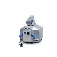
| O2k | O2k | O2k - Oroboros O2k: the modular system for high-resolution respirometry. |
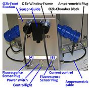
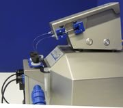
| O2k-Fluo LED2-Module | The O2k-Fluo LED2-Module is a component of the O2k-Fluorometer (O2k-Series D to G). It is an amperometric add-on module to the O2k-Core (O2k-Series D to G), adding a new dimension to high-resolution respirometry. Optical sensors are inserted through the front window of the O2k-glass chambers, for measurement of hydrogen peroxide production (Amplex® UltraRed), ATP production (Magnesium Green™), mt-membrane potential (Safranin, TMRM, Rhodamine 123), Ca2+ (Calcium Green™), and numerous other applications open for O2k-user innovation. | |
| O2k-Fluo Smart-Module | The O2k-Fluo Smart-Module is an amperometric add-on module to the O2k-Respirometer, adding a new dimension to high-resolution respirometry. Optical sensors are inserted through the front window of the O2k-glass chambers, for measurement of hydrogen peroxide production (Amplex® UltraRed), ATP production (Magnesium Green™), mt-membrane potential (Safranin, TMRM), Ca2+ (Calcium Green™), and numerous other applications open for O2k-user innovation. | |
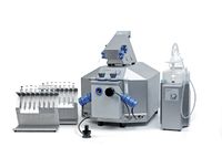
| O2k-FluoRespirometer | The Oroboros O2k-FluoRespirometer - the experimental system complete for high-resolution respirometry (HRR), including fluorometry, the TIP2k and the O2k-sV-Module allowing simultaneous monitoring of oxygen consumption together with either ROS production (AmR), mt-membrane potential (TMRM, Safranin and Rhodamine 123), Ca2+ (CaG) or ATP production (MgG). The O2k-FluoRespirometer supports all add-on O2k-Modules: O2k-TPP+ ISE-Module, O2k-pH ISE-Module, O2k-NO Amp-Module, enabling measurement of mt-membrane potential with ion sensitive electrodes (ISE for TPP+ or TPMP+) or pH. | |
| O2k-sV-Module | O2k-sV-Module | The O2k-sV-Module is the O2k small-volume module, comprised of two Duran® glass chambers of 12 mm inner diameter specifically developed to perform high-resolution respirometry with reduced amounts of biological sample, and all the components necessary for a smaller operation volume V of 0.5 mL. The current DatLab version is included in the delivery of this revolutionary module. |
| Optics | Optics are the components that are used to relay and focus light through a spectrofluorometer or spectrophotometer. These would normally consist of lenses and/or concave mirrors. The number of such components should be kept to a minimum due to the losses of light (5-10%) that occur at each surface. | |
| PH calibration buffers | pH calibration buffers are prepared to obtain two or more defined pH values for calibration of pH electrodes and pH indicator dyes. | |
| Phosphorescence | Phosphorescence is a similar phenomenon to fluorescence. However, instead of the electron returning to its original energy state following excitation, it decays to an intermediate state (with a different spin value) where it can remain for some time (minutes or even hours) before decaying to its original state. Phosphorescence is one form of Luminescence, especially Photoluminescence. | |
| Photodiode arrays | Photodiode arrays are two dimensional assemblies of photodiodes. They are frequently used in conjunction with charge coupled devices (CCDs) for digital imaging. They can be used in combination with dispersion devices to detect wavelength dependent light intensities in a spectrofluorometer or spectrophotometer. | |
| Photodiodes | Photodiodes are photodetectors that convert incident light into a current or voltage dependent on their configuration. They have replaced photomultiplier tubes for most applications. For fluorometric measurements that do not require spectral data, a single photodiode with suitable filters can be used. Due to their larger detection area, they are more sensitive than photodiode arrays. | |
| Polyether ether ketone | PEEK | Polyether ether ketone (PEEK) is a semicrystalline organic polymer thermoplastic, which is chemically very resistant, with excellent mechanical properties. PEEK is compatible with ultra-high vacuum applications, and its resistance against oxygen diffusion make it an ideal material for high-resolution respirometry (POS insulation; coating of stirrer bars; stoppers for closing the O2k-Chamber). |
| Power O2k-FluoRespirometer | Power O2k-FluoRespirometer - optional configuration as additional system for increasing output combined with the O2k-FluoRespirometer (O2k-Series H). The Power O2k-FluoRespirometer includes the TIP2k and the O2k-sV-Module, and supports all add-on O2k-Modules of the Oroboros O2k. It can be added to an existing Oroboros O2k of any O2k-Series. This application does not require an additional ISS-Integrated Suction System and O2k-Titration Set. Furthermore, the OroboPOS-Mounting Tool of the OroboPOS Service Tools can be used from the available O2k and is not included. | |
| Quenching | Quenching is the name given to any process that reduces fluorescence intensity. Molecular oxygen is a fluorescence and phosphorescence quencher for some substances – a phenomenon that has been made use of in constructing optical probes for measuring oxygen. | |
| Reactive oxygen species | ROS | Reactive oxygen species, ROS, are molecules derived from molecular oxygen, including free oxygen radicals, which are more reactive than O2. Physiologically and pathologically important ROS include superoxide, the hydroxyl radical and hydroxide ion, hydrogen peroxide and other peroxides. These are important in cell signalling, oxidative defence mechanisms and oxidative stress. |
| Resolution | Spectral resolution is a measure of the ability of an instrument to differentiate between two adjacent wavelengths. Two wavelengths are normally considered to be resolved if the minimum detector output signal (trough) between the two peaks is lower than 80 % of the maximum. The resolution of a spectrofluorometer or spectrophotometer is dependent on its bandwidth. | |
| Resorufin | Res | Resorufin is a fluorescence probe used in various biological assays. Among others, it is the product obtained in the Horseradish peroxidase-catalyzed assay using Amplex Red for the measurement of H2O2 production. |
| Rhodamine 123 | Rh123 | Rhodamine 123 (Rh123) is an extrinsic fluorophore and can be used as a probe to determine changes in mitochondrial membrane potential. Rh123 is a lipophilic cation that is accumulated by mitochondria in proportion to Δψmt. Using ethanol as the solvent, the excitation maximum is 511 nm and the emission maximum is 534 nm. The recommended excitation and emission wavelengths in PBS are 488 and 515-575 nm, respectively (Sigma-Aldrich). |
| SUIT-003 AmR ce D017 | CCP-ce S permeability test | 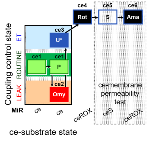 |
| SUIT-006 | CCP-mtprep | 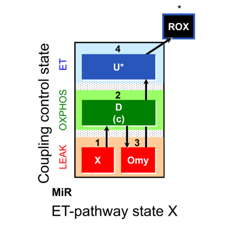 |
| SUIT-006 AmR mt D048 | CCP mt PM - AmR | 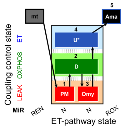 |
| SUIT-006 Fluo mt D034 | CCP mt PM - Fluo | 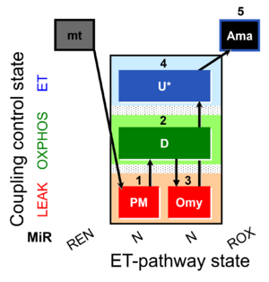 |
| SUIT-006 MgG ce-pce D085 | CCP MgG ce-pce | 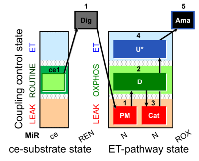 |
| SUIT-006 MgG mt D055 | CCP MgG mt | 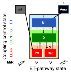 |
| SUIT-009 | 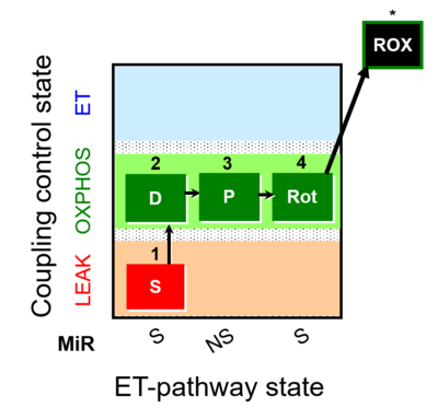 | |
| SUIT-009 AmR ce-pce D019 | H2O2 RET ce-pce S_L | 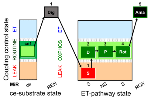 |
| SUIT-009 AmR mt D021 | H2O2 mtprep | 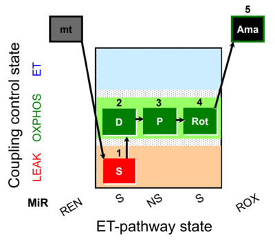 |
| SUIT-013 | ce | 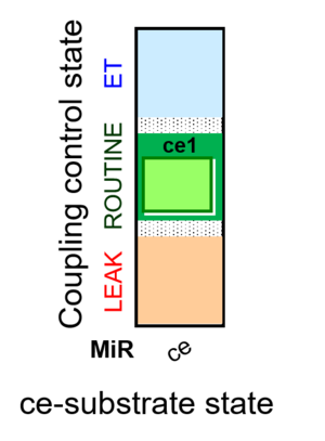 |
| SUIT-013 AmR ce D023 | O2 dependence of H2O2 production ce | 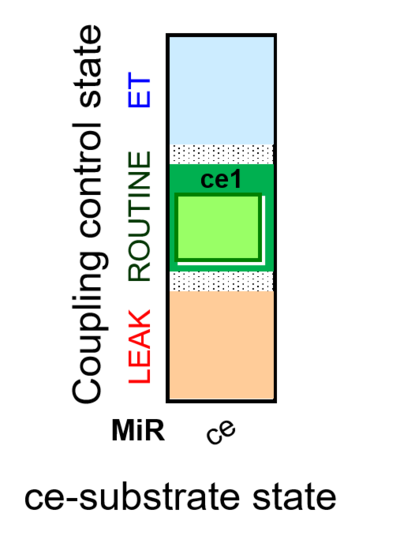 |
| SUIT-018 | O2 dependence-H2O2 | 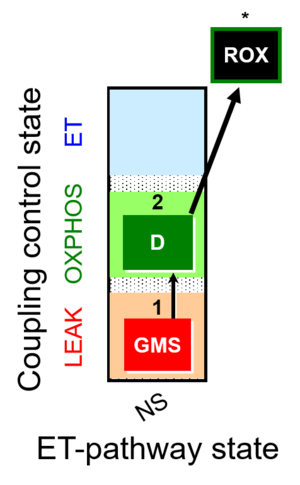 |
| SUIT-018 AmR ce-pce D068 | O2 dependence-H2O2 | 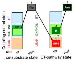 |
| SUIT-018 AmR mt D031 | O2 dependence-H2O2 | 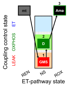 |
| SUIT-018 AmR mt D040 | NS(GM) |  |
| SUIT-018 AmR mt D041 | O2 dependence-H2O2 |  |
| SUIT-020 | PM+G+S+Rot_OXPHOS+Omy | 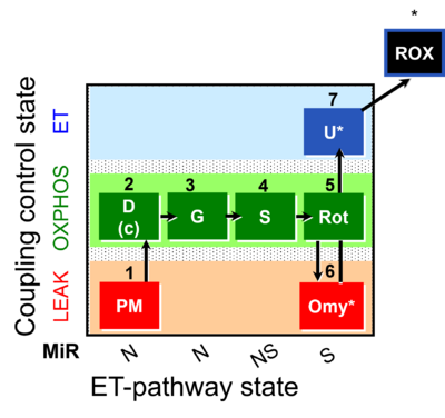 |
| SUIT-020 Fluo mt D033 | NS(PGM) | 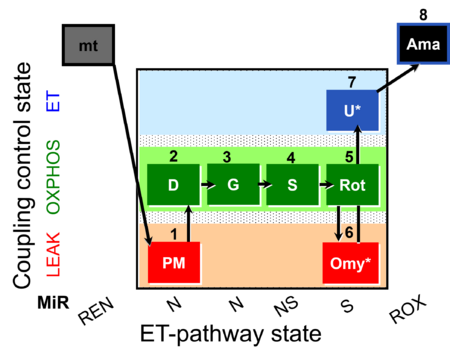 |
| SUIT-021 | OXPHOS (GM+S+Rot+Omy) | 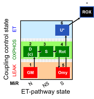 |
| SUIT-021 Fluo mt D036 | NS(GM) | 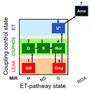 |
| SUIT-026 | RET | 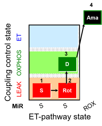 |
| SUIT-026 AmR ce-pce D087 | RET | 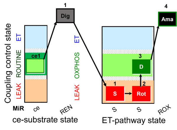 |
| SUIT-026 AmR mt D064 | RET | 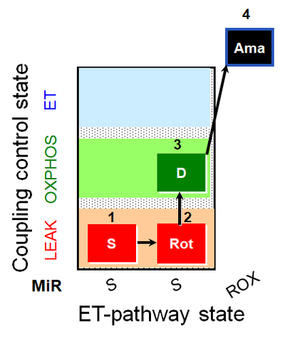 |
| SUIT-026 AmR mt D077 | RET |  |
| SUIT-026 O2 ce-pce D088 | RET (respiratory control) of SUIT-026 AmR ce-pce D087 | 400px |
| SUIT-026 O2 mt D063 | RET (respiratory control) of SUIT-026 AmR mt D064 | 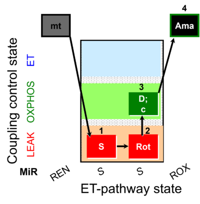 |
| SUIT-027 O2 ce-pce D065 | Malate anaplerosis | 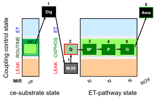 |
| SUITbrowser | Use the SUITbrowser to find the substrate-uncoupler-inhibitor-titration (SUIT) protocol most suitable for addressing your research questions.
Open the SUITbrowser: http://suitbrowser.oroboros.at/
 How to find a DL-Protocol (DLP) How to find a DL-Protocol (DLP) | |
| Safranin | Saf | Safranin is one of the most established dyes for measuring mitochondrial membrane potential by fluorometry. It is an extrinsic fluorophore with an excitation wavelength of 495 nm and emission wavelength of 587 nm. Safranin is a potent inhibitor of N-linked respiration and of the phosphorylation system. Synonyms: Safranin O, Safranin Y, Safranin T, Gossypimine, Cotton Red, Basic Red2 |
| Scattering | Most biological samples do not consist simply of pigments but also particles (e.g. cells, fibres, mitochondria) which scatter the incident light. The effect of scattering is an apparent increase in absorbance due to an increase in pathlength and the loss of light scattered in directions other than that of the detector. Two types of scattering are encountered. For incident light of wavelength λ, Rayleigh scattering is due to particles of diameter < λ (molecules, sub-cellular particles). The intensity of scatter light is proportional to λ4 and is predominantly backward scattering. Mie scattering is caused by particles of diameter of the order of or greater than λ (tissue cells). The intensity of scatter light is proportional to 1/λ and is predominantly forward scattering. | |
| Selectivity | Selectivity is the ability of a sensor or method to quantify accurately and specifically the analyte or analytes in the presence of other compounds. | |
| Sensitivity | Sensitivity refers to the response obtained for a given amount of analyte and is often denoted by two factors: the limit of detection and the limit of quantification. | |
| Signal-to-noise ratio | S/N | The signal to noise ratio is the ratio of the power of the signal to that of the noise. For example, in fluorimetry it would be the ratio of the square of the fluorescence intensity to the square of the intensity of the background noise. |
| Slit width | The slit width determines the amount of light entering the spectrofluorometer or spectrophotometer. A larger slit reduces the signal-to-noise ratio but reduces the wavelength resolution. | |
| Smoothing | Various methods of smoothing can be applied to improve the signal-to-noise ratio. For instance, data points recorded over time [s] or over a range of wavelengths [nm] can be smoothed by averaging n data points per interval. Then the average of the n points per smoothing interval can be taken for each successively recorded data point across the time range or range of the spectrum to give a n-point moving average smoothing. This method decreases the noise of the signal, but clearly reduces the time or wavelength resolution. More advanced methods of smoothing are applied to retain a higher time resolution or wavelength resolution. | |
| Spectrofluorometer | A spectrofluorometer makes use of a spectrometer to measure and analyse the fluorescent emission spectra from a fluorophore. It will typically differ from an absorbance spectrophotometer in that it will have a larger slit width (to increase sensitivity) and use a longer integration time. The configuration of the illuminating and receiving optics also differ from spectrophotometry in that the excitation source is directed perpendicularly to the position of the emission detector so that the intensity of the excitation signal reaching the detector is minimised. | |
| Spectroscopy | Spectroscopy is a broader term than spectrophotometry in that it is concerned with the investigation and measurement of spectra produced when matter interacts with or emits any form electromagnetic radiation. | |
| Spline | Some spectrofluorometer or spectrophotometer software offers the possibility of spline interpolation of the spectral data points. This makes use of a polynomial (the number of spline points is entered by the user) to interpolate the curve between the data points. | |
| Stability | Stability determines the accuracy of intensity and absorbance measurements as a function of time. Instability (see drift introduces systematic errors in the accuracy of fluorescence and absorbance measurements. | |
| Steady state | A system is in a steady state if the state variables of a dynamic system do not change over time due to exchange processes with the environment, which compensate for internal dissipative transformations — such as chemical reactions or diffusion — and thus prevent any changes of the system and externalize dissipative changes to the environment. The dynamic nature of the steady state differentiates it from the thermodynamic equilibrium state. {Quote} Steady states can be obtained only in open systems, in which changes by internal transformations, e.g., O2 consumption, are instantaneously compensated for by external fluxes across the system boundary, e.g., O2 supply, thus preventing a change of O2 concentration in the system (Gnaiger 1993). Mitochondrial respiratory states monitored in closed systems satisfy the criteria of pseudo-steady states for limited periods of time, when changes in the system (concentrations of O2, fuel substrates, ADP, Pi, H+) do not exert significant effects on metabolic fluxes (respiration, phosphorylation). Such pseudo-steady states require respiratory media with sufficient buffering capacity and substrates maintained at kinetically-saturating concentrations, and thus depend on the kinetics of the processes under investigation. {end of Quote: BEC 2020.1}. Whereas fluxes may change at a steady state over time, concentrations are maintained constant. The 'respiratory steady state' (Chance and Williams 1955) is characterized by constant fluxes (O2 flux, H2O2 flux) and measured variables of state (cytochrome redox states, Q redox state, NADH redox state, mitochondrial membrane potential). High-resolution respirometry allows for the measurement of several parameters (e.g. O2 flux, H2O2 flux, mitochondrial membrane potential) at pseudo-steady states, when changes of concentrations in the closed system do not exert any control on fluxes. Combination with the Titration-Injection microPump (TIP2k) allows operation with programmable titration regimes at steady-state ADP concentration (Gnaiger 2001), oxygen concentration (oxystat mode; Gnaiger et al 2000, Harrison et al 2015) or steady-state pH (pH-stat more), yielding an expanded flexibility in experimental design by combining the technical advantages of closed and open systems approaches. | |
| Stray light | Stray light is defined as the detected light of any wavelength that lies outside the bandwidth of the selected wavelength. In the presence of stray light of intensity Is, the equation for transmittance (T) becomes T = (I + Is)/(I0 + Is) where I0 is the incident light intensity and I is the transmitted light intensity. Clearly, the lower the value of I, the more dominant becomes the stray light term and so can cause errors in the quantification of low fluorescence signals or at high levels of absorbance. | |
| TIP2k-Module | TIP2k-Module - Titration-Injection microPump (TIP2k) for two-channel operation with the O2k-FluoRespirometer with automatic control by DatLab of programmable titration regimes and feedback control (oxystat, pH-stat). | |
| TMRM | TMRM | TMRM (tetramethylrhodamine methyl ester) is an extrinsic fluorophore used as a probe to determine changes in mitochondrial membrane potential. TMRM is a lipophilic cation that is accumulated in the mitochondrial matrix in proportion to Δψmt. Upon accumulation of the dye it exhibits a red shift in its absorption and fluorescence emission spectrum. The fluorescence intensity is quenched when the dye is accumulated in the mitochondrial matrix. |
| Taurine | Taurine, or 2-Aminoethan sulfonic acid, is one of the most abundant low-molecular-weight organic constituents in animals and humans. It has a multitude of functions in different types of tissue, one of which is the stabilization of membranes. Because of this and its antioxidative effect, taurine is a component of the respiration media MiR05 and MiR06 to preserve mitochondrial function. | |
| Wavelength averaging | Wavelength averaging is the averaging of several adjacent data points across the recorded spectrum (spectral smoothing), to improve the signal-to-noise ratio. For example, if the instrument recorded 5 data points per nm, the average of the 5 points can be taken for each successive nm across the range of the spectrum to give a 5-point smoothing. This method clearly reduces the wavelength resolution. |

