Nogueras 2017 MiPschool Obergurgl
| Mitochondrial dysfunction in peripheral blood mononuclear cells from multiple sclerosis patients. |
Link: MitoEAGLE
Nogueras L, Gil A, Hervas JV, Gonzalez C, Martin-Gari M, Canudes M, Peralta S, Moga MJ, Boada J, Pamplona R, Portero M, Brieva L, Gonzalo H (2017)
Event: MiPschool Obergurgl 2017
Multiple sclerosis (MS) is a chronic, inflammatory, and demyelinating disease of the central nervous system (CNS). Its etiology is still unknown; nevertheless, the abundance of immune cells in CNS lesions of MS patients supports the notion that MS is an immune-mediated disorder[1,2]. On the other hand mitochondrial dysfunction might have a central role in the pathophysiology of MS. Different studies have reported a mitochondrial impairment in the CNS but the results in the immune system are still inconsistent[3].
Cellular immune function depends on energy supply and mitochondrial function that is why this study has investigated mitochondrial respiration (Oroboros O2k; Oroboros Instruments, Innsbruck, Austria) in peripheral blood mononuclear cells (PBMCs), an established model to investigate the mitochondrial impairments. Mitochondrial respiration was assessed in intact PBMCs in 45 individuals with a diagnosis of MS compared with 29 healthy age gender-matched controls using high-resolution respirometry. Furthermore we analyzed superoxide production of lymphocytes (they represent between 70-90% of PBMCs cells) by median fluorescent intensity (MFI) of MitoSoxTM (Red mitocondrial superoxide indicator, Molecular Probes, Invitrogen, Barcelona, Spain) by flow citometry.
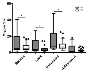
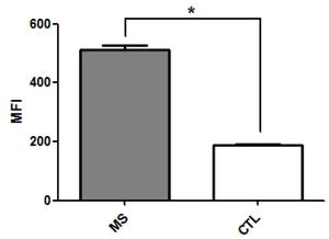
Individuals with MS showed a significant increase in ROUTINE respiration (p=0.045), uncoupled respiration (after oligomycin addition p=0.035), as well as in the maximum respiratory capacity (after carbonyl cyanide-p-trifluoromethoxyphenylhydrazone titration p=0.035). Finally Antimycin A was added but not significant differences were founded between groups (Figure 1). In addition, PBMCs of these patients produced more superoxide than those in control group individuals (p=0.021) (Figure 2).
PBMCs cells in MS patients show a significant increase of uncoupled respiration which is consistent with immune system activation and with an autoimmune disease. A higher maximal uncoupled respiration can indicate a higher capacity of mitochondrial respiration and is related to superoxide production which is also higher in MS patients[4]. In conclusion, our results support systemic involvement in MS, and reveal the study of mitochondrial function in blood cells as a source of potential biomarkers of the disease.
• Bioblast editor: Kandolf G
• O2k-Network Lab: ES Lleida Boada J
Labels: MiParea: Respiration, Patients Pathology: Neurodegenerative
Tissue;cell: Blood cells
Preparation: Intact cells
Coupling state: LEAK, ROUTINE, ET
Pathway: ROX
HRR: Oxygraph-2k
Event: C1, Oral
PMBCs
Affiliations
- Nogueras L(2), Gil A(2), Hervás JV(3), Gonzalez C(3), Martin-Gari M(2), Canudes M(2), Peralta S(1), Moga MJ(3), Boada J(2), Pamplona R(2), Portero M(2), Brieva L(3), Gonzalo H(1,3)
- Fundación Esclerosis Multiple
- Inst recerca biomèdica de Lleida
- Arnau Vilanova Hospital
- Lleida, Spain. - lara.noguerasp@gmail.com
Figures
Figure 1. Boxplots of group-wise comparison of respiration in PBMCs from MS patients (n = 45) and control subjects (n = 29) characterized by mitochondrial oxygen flux.
Asterisk (*) indicates significant group differences on an alpha level of 0.05 (Student’s independent ttest).
Leak: flux oxygen after oligomycin addition
Uncoupled: flux oxygen after carbonyl cyanide-p-trifluoromethoxyphenylhydrazone titration
Figure 2. Boxplots of group-wise comparison of superoxide production of lymphocytes from MS patients (n = 45) and control subjects (n = 29) characterized by median fluorescent intensity.
Asterisk (*) indicates significant group differences on an alpha level of 0.05 (Student’s independent ttest).
MFI: median fluorescent intensity
References and support
- Legroux L, Arbour N (2015) Multiple sclerosis and T lymphocytes: an entangled story. J Neuroimmune Pharmacol 10:528-46.
- Haghikia A, Faissner S, Pappas D, Pula B, Akkad DA, Arning L, Ruhrmann S, Duscha A, Gold R, Baranzini SE, Malhotra S, Montalban X, Comabella M, Chan A (2015) Interferon-beta affects mitochondrial activity in CD4+ lymphocytes: Implications for mechanism of action in multiple sclerosis. Mult Scler 21:1262–70.
- Petzold A, Nijland PG, Balk LJ, Amorini AM, Lazzarino G, Wattjes MP, Gasperini C, van der Valk P, Tavazzi B, Lazzarino G, van Horssen J (2015) “Visual pathway neurodegeneration winged by mitochondrial dysfunction. Ann Clin Transl Neurol 2:140–50.
- Karabatsiakis A, Böck C, Salinas-Manrique J, Kolassa S, Calzia E, Dietrich DE, Kolassa IT (2014) Mitochondrial respiration in peripheral blood mononuclear cells correlates with depressive subsymptoms and severity of major depression. Transl Psychiatry 4:e397.
- Selected mentor: Jordi Boada


